Am I a Candidate for Kyphoplasty?

Bone is living tissue that continually remodels itself as cells grow, dies off, and new cells grow in their place. Osteoporosis is a bone disease that develops when cell breakdown exceeds cell production. The bones lose mass and density, becoming weak and brittle. They can become so weak that you can break them just from sneezing. Spontaneous compression fractures in the spine are a hallmark of the disease.
At Vertrae®, board-certified neurosurgeon Dr. Kamal R. Woods and his team treat all manner of spinal conditions, including osteoporosis-caused compression fractures, through spinal surgery, minimally invasive spinal surgery, and robotic surgery. The procedure used for compression fractures is known as kyphoplasty. Here’s what’s involved.
Osteoporosis revealed
If you look at healthy bone tissue under a microscope, it looks much like a honeycomb, with thick walls and evenly spaced holes. Osteoporosis means “porous bone.” When you look at bone tissue from a patient with the disease, the walls are thin, and the holes are large and irregularly spaced due to tissue loss.
Because porous bone has more space than tissue, the bones break more easily than a healthy bone would. Anyone can develop the disease, but those at greatest risk from non-controllable causes (i.e. genetics, race) are small, white or Asian, postmenopausal women.
Osteoporosis is also called a “silent disease” because you have no way to tell if your bones aren’t healthy without a bone density scan, usually a dual-energy X-ray absorptiometry (DXA) (pronounced DEXA) scan.
Signs you have the disease are breaking bones easily, especially in the hip and spine, losing height, and/or developing upper back curvature. If you experience any of these, contact Vertrae® as soon as possible. An accurate diagnosis can lead to appropriate and effective treatment.
What is a compression fracture?
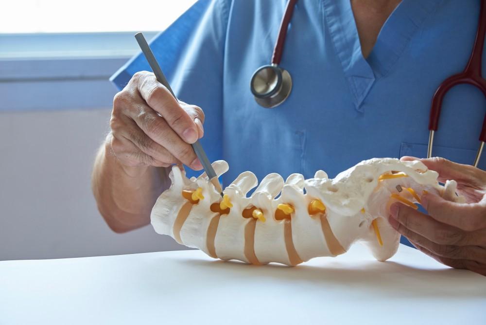
Your spine contains 24 bony vertebrae joined together by facet joints, and each pair has a cushiony disc between the bones that absorbs shock when you walk, jump, or turn.
If your vertebrae lose density, your upper spine may start to curve with the stress. This can cause a humpback, or the vertebrae may collapse into each other — a compression fracture. These extremely painful fractures may prevent you from doing the most basic things, such as bending over or getting out of bed.
Compression fractures generally occur in the thoracic (chest) region of the spine, which contains the T1-T12 vertebrae; however, they can also occur in the lumbar (lower back) spine, containing vertebrae L1-L5.
Am I a good candidate for kyphoplasty?
Kyphoplasty can also be used as part of a patient’s cancer treatment. Still, vertebral compression fractures caused by osteoporosis tend to respond better to kyphoplasty (and vertebroplasty) than those caused by cancer. Kyphoplasty complication rates may be more than twice as high when the vertebral compression fracture is caused by cancer (10%) compared to osteoporosis (4%).
When the compression fracture occurs at the front of the vertebral body, it’s called a wedge fracture. Wedge fractures are the most common compression fractures and are the type that responds best to kyphoplasty or vertebroplasty treatments. Other fracture shapes, including biconcave fractures (front and back are intact, but the middle part’s compressed) or crush fractures (entire vertebral body breaks), aren’t as likely to have positive outcomes.
As a result, the best candidate is one with a wedge-shaped compression fracture in a vertebral body. Patients who have other conditions like disc herniation, arthritis, or stenosis aren’t good candidates for this procedure, but they may be helped with other surgeries.
Kyphoplasty for compression fractures
Dr. Woods uses a minimally invasive approach to kyphoplasty, with the goals of stabilizing the fractured vertebra(e), restoring the vertebrae to their normal height, and relieving the pain from the break. He uses an inflated balloon to establish a cavity inside the vertebral body, filling it with PMMA, a surgical cement. The procedure takes about an hour per vertebra.
Once you're home, you can return to your normal activities, though we recommend avoiding strenuous activities like lifting, driving, and intense exercise for at least six weeks. Some patients feel immediate pain relief from the procedure, while others need a day or so before their pain level drops.
If you have painful compression fractures or are at risk of developing them, it’s time to come to Vertrae® for an evaluation with Dr. Woods. To schedule, call our office at 844-255-2225 or book online today.
You Might Also Enjoy...


4 Benefits of Outpatient Spine Surgery

Pulled Muscle vs. Pinched Nerve: What's the Difference?

4 Subtle Signs of Sciatica

When to Consider Back Surgery for a Herniated Disc

