A Closer Look at Your Sacroiliac Joint
Your sacroiliac (SI) joints connect your spine to your hip bones while supporting movement and bearing the upper body's weight. Learn more about the SI joints and how they work.

It used to be that all surgeries were done with an “open” technique that involved long incisions, extended hospital stays and recovery times, and increased risks of blood loss and infection.
In the 1980s, minimally invasive surgery emerged as a safe and effective technique for many procedures. One of the first types employed was laparoscopy — surgery done through one or more small incisions that used small tubes and tiny cameras and surgical instruments threaded through them to reach the area that needed observation and/or repair.
The idea of robotics for surgical assistance was proposed in 1967, but actual use didn’t start until the late 1980s with the Robodoc from Integrated Surgical Systems in Sacramento, California. It was an orthopedic image-guided system used in prosthetic hip replacement. Robot-assisted surgery platforms provide a magnified 3D view of the treatment area and give the surgeon greater precision and control of the technique.
Although now multipurpose robotic systems are ubiquitous for minimally invasive surgery, early prototypes were specialty-focused and originally intended for long-distance trauma surgery in battlefield settings.
While Dr. Kamal R. Woods and his team at Vertrae® often use conservative methods to treat spinal problems, some conditions warrant surgery. The team specializes in using minimally invasive techniques, including the use of robot-assisted surgery. Here’s what you need to understand about the different types of minimally invasive surgery and how they can address problems with your spine.
Along with large incisions, open spine surgery depended on the surgeon cutting through layers of muscle and connective tissue to reach the spine and repair it. It was very invasive and left the patient with long recovery times because of the damaged tissue.
But if minimally invasive techniques use only buttonhole-sized incisions, how can the doctor reach the spine and place any hardware that’s needed to strengthen and support it? The answer lies in the tubular retractor.
Inserted into the soft tissue, the retractor progressively dilates, widening the access space to the spine. Sometimes, the surgeon also uses an endoscope or microscope focused down the tube to provide a better view through the small incision. Once the procedure is complete, he removes the retractor, and the dilated tissues come back together.
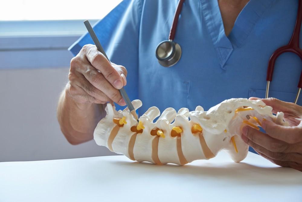
Traditionally, placing rods, plates, and screws required removing large amounts of muscle and other tissues from the spine’s surface. In minimally invasive techniques, the surgeon places them percutaneously — through the skin without tissue dissection.
Aided by X-ray images, the surgeon threads guidewires through the skin and into the spinal vertebrae along the path the screws need to travel. Next, he places the screws over the guidewires. The screws have temporary extenders that reach outside the skin and help guide the passage of rods to connect and secure the screws. Finally, the surgeon removes the extenders and closes the wound.
Spinal instrumentation placement is one area that has benefited tremendously from robot-assisted surgery, allowing the hardware to be placed more safely and accurately.
MIS can treat many different spinal conditions, including:
The procedures relieve the pain these conditions cause and restore function to the spine.
Some of the MIS procedures Dr. Woods uses include:
In a laminectomy, Dr. Woods removes the back part of the vertebra that covers and protects the spinal canal (lamina). This reduces painful pressure on the spinal cord or a spinal nerve due to lumbar stenosis, a herniated disc, or a bone spur (bony growth on the vertebra).
If a spinal disc bulges or herniates, the disc material can compress nearby nerves, causing pain. Here, Dr. Woods trims or removes the herniated disc, using tubular dilators and a microscope to access the spine.
Spinal stenosis can lead to pain, numbness, and muscle weakness by compressing nerve roots. This procedure removes the bone and soft tissues causing the compression, once again using tubular dilators and a microscope to access the damaged area.
Dr. Woods uses a bone graft to fuse two adjacent vertebrae in your spine if you’ve developed spinal instability due to conditions like degenerative disc disease and disc herniation. Fusing the vertebrae leads to increased stability because the spine can no longer move at that level.
If you’d like to learn more about the different types of minimally invasive surgery, or if you’re dealing with back pain and stiffness and need a diagnosis and treatment, call Vertrae at either our Dayton or Springfield location to set up a consultation with Dr. Woods, or book online with us today.